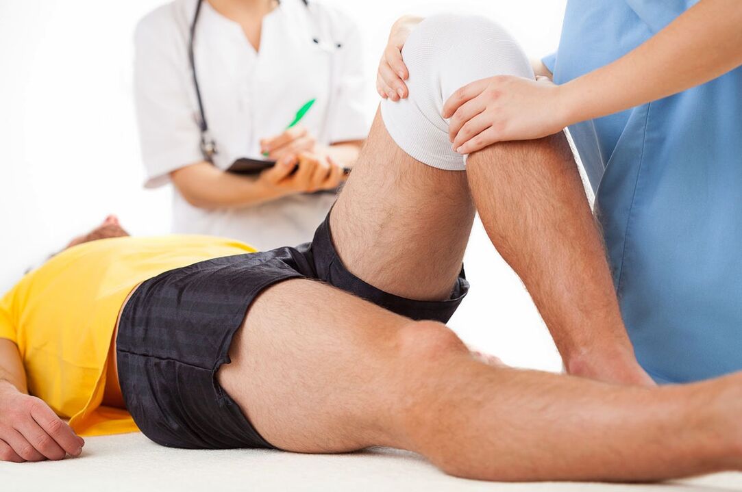
Arthrosis is the most common joint disease. According to experts, 6. 43% of the population of our country suffers from it. Men and women suffer from arthrosis equally often, however, among young patients there is a slight predominance of men, and among the elderly - women. An exception to the general picture is arthrosis of the interphalangeal joints, which develops in women 10 times more often than in men.
With age, the incidence increases dramatically. So, according to research, arthrosis is detected in 2% of people younger than 45 years old, in 30% of people from 45 to 64 years old and in 65-85% in people aged 65 years and older. Arthrosis of the knee, hip, shoulder and ankle joints is of the greatest clinical significance due to its negative impact on the standard of living and working capacity of patients.
Causes
In some cases, the disease occurs for no apparent reason, such arthrosis is called idiopathic or primary.
There is also a secondary arthrosis - developed as a result of some pathological process. The most common causes of secondary arthrosis are:
- Injuries (fractures, meniscus injuries, ligament ruptures, dislocations, etc. ).
- Dysplasia (congenital joint developmental disorders).
- Degenerative-dystrophic processes (Perthes disease, osteochondritis dissecans).
- Diseases and conditions in which there is increased mobility of the joints and weakness of the ligamentous apparatus.
- Hemophilia (arthrosis develops as a result of frequent hemarthrosis).
Risk factors for the development of arthrosis include:
- Elderly age.
- Overweight
- Excessive stress on the joints or a specific joint.
- Surgical interventions on the joint,
- Hereditary predisposition (the presence of arthrosis in the next of kin).
- Endocrine imbalance in postmenopausal women.
- Neurodystrophic disorders in the cervical or lumbar spine (shoulder arthritis, lumbar-iliac muscle syndrome).
- Repetitive joint microtrauma.
Pathogenesis
Arthrosis is a polyetiological disease, which, regardless of the specific causes of its occurrence, is based on a violation of the normal formation and restoration of cartilaginous tissue cells.
Normally, the articular cartilage is smooth and elastic. This allows the articular surfaces to move freely relative to each other, provides the necessary shock absorption and, thereby, reduces the load on the adjacent structures (bones, ligaments, muscles and capsule). With arthrosis, the cartilage becomes rough, the articular surfaces begin to "cling" to each other during movement. The cartilage looses more and more. Small pieces are separated from it, which fall into the joint cavity and move freely in the joint fluid, injuring the synovium. In the superficial zones of the cartilage, small foci of calcification appear. In the deep layers, areas of ossification appear. In the central zone, cysts are formed, communicating with the joint cavity, around which, due to the pressure of the intra-articular fluid, ossification zones are also formed.
Pain syndrome
Pain is the most constant symptom of arthrosis. The most striking signs of pain in arthrosis are the connection with physical activity and with the weather, night pains, starting pain and sudden sharp pains in combination with joint blockade. With prolonged exertion (walking, running, standing), the pain intensifies, and at rest they subside. The cause of nocturnal pain in arthrosis is venous congestion, as well as an increase in intraosseous blood pressure. The pains are aggravated by unfavorable weather factors: high humidity, low temperature and high atmospheric pressure.
The most characteristic sign of arthrosis is starting pain - pain that occurs during the first movements after a state of rest and disappears while maintaining motor activity.
Symptoms
Arthrosis develops gradually, gradually. Initially, patients are worried about mild, short-term pain without a clear localization, aggravated by physical exertion. In some cases, the first symptom is crunching when moving. Many patients with arthrosis report a feeling of discomfort in the joint and transient stiffness during the first movements after a period of rest. Subsequently, the clinical picture is complemented by night and weather pains. Over time, the pain becomes more and more pronounced, there is a noticeable restriction of movement. Due to the increased load, the joint on the opposite side begins to hurt.
The periods of exacerbations alternate with remissions. Exacerbations of arthrosis often occur against a background of increased stress. Because of the pain, the muscles of the limbs reflexively spasm, and muscle contractures can form. The crunch in the joint becomes more and more constant. At rest, muscle cramps and discomfort in muscles and joints appear. Due to the growing deformation of the joint and severe pain syndrome, lameness occurs. In the later stages of arthrosis, the deformity becomes even more pronounced, the joint is bent, movements in it are significantly limited or absent. Support is difficult; when moving, a patient with arthrosis has to use a cane or crutches.
Diagnostics
The diagnosis is made on the basis of characteristic clinical signs and X-ray picture of arthrosis. X-rays are taken of the diseased joint (usually in two projections): with gonarthrosis - X-ray of the knee joint, with coxarthrosis - X-ray of the hip joint, etc. The X-ray picture of arthrosis consists of signs of dystrophic changes in the area of articular cartilage and adjacent bone. The joint gap is narrowed, the bone site is deformed and flattened, cystic formations, subchondral osteosclerosis and osteophytes are revealed. In some cases, with arthrosis, signs of joint instability are found: curvature of the axis of the limb, subluxation.
Taking into account the radiological signs, specialists in the field of orthopedics and traumatology distinguish the following stages of arthrosis (Kellgren-Lawrence classification):
- Stage 1 (doubtful arthrosis) - a suspicion of narrowing of the joint space, osteophytes are absent or present in small numbers.
- Stage 2 (mild arthrosis) - suspicion of narrowing of the joint space, osteophytes are clearly defined.
- Stage 3 (moderate arthrosis) - a clear narrowing of the joint space, there are clearly pronounced osteophytes, bone deformities are possible.
- Stage 4 (severe arthrosis) - pronounced narrowing of the joint space, large osteophytes, pronounced bone deformities and osteosclerosis.
Sometimes X-rays are not enough to accurately assess the condition of the joint. To study bone structures, CT of the joint is performed, to assess the state of soft tissues - MRI of the joint.
Treatment
The main goal of treating patients with arthrosis is to prevent further cartilage destruction and to preserve the function of the joint.
During the period of remission, a patient with arthrosis is sent to physical therapy. The set of exercises depends on the stage of arthrosis.
Drug treatment in the phase of exacerbation of arthrosis includes the appointment of non-steroidal anti-inflammatory drugs, sometimes in combination with sedatives and muscle relaxants.
Long-term use of arthrosis includes chondroprotectors and synovial fluid prostheses.
To relieve pain, reduce inflammation, improve microcirculation and eliminate muscle spasms, a patient with arthrosis is referred for physiotherapy. In the exacerbation phase, laser therapy, magnetic fields and ultraviolet irradiation are prescribed, in the remission phase - electrophoresis with dimexide, trimecaine or novocaine, phonophoresis with hydrocortisone, inductothermy, thermal procedures (ozokerite, paraffin), sulfide, radon and sea baths. Electrical stimulation is performed to strengthen the muscles.
In case of destruction of articular surfaces with pronounced dysfunction of the joint, arthroplasty is performed.


